Life Processes Class 10 Notes | Nutrition, Respiration, Transportation, Excretion | Easy Explanation
How do we know that plant/animal is living?
- Plants are green and fresh.
- They grow.
- But we cannot see the visible growth and visible breathing in animals.
- visible movement is not characteristic of life.
Why are molecular movements needed for life?
- We know that all the living organisms are in a well-organised structure.
- cells → tissues → organs → organ systems
- Due to environment this organised structure sometimes are breaking down over time to time.
- So they keep on repairing and maintaining the structure.
- Since these structures are made up of molecules, molecular level repair and maintenance takes place.
Why maintenance of life process is needed?
Maintenance of life process is needed to prevent damage.
What are the processes essential for maintaining life?
- Respiration
- Digestion
- Circulation
- Transportation
- Excretion
What are the outside raw materials used for by an organism?
- The outside raw materials used is food and O₂.
- Raw material requirement varies depending on the complexity of organism, & its environment (where it lives)
Where do plants get each of the raw material required for photosynthesis?
- CO₂ from atmosphere through stomata
- water absorbed by plant roots from soil.
- Sunlight
- Other nutrients obtained by soil by plant roots.
What criteria do we use to decide whether something is alive?
- walking
- Breathing → visible changes used to determine whether something is alive or dead.
- Growth
However, some organism will not show visible changes.
Why diffusion insufficient to meet the O₂ requirement of multicellular organisms?
- Multicellular organism require lot of O₂ to diffuse into the body quickly inorder to meet the O₂ requirement.
- Diffusion is a slow process and takes lot of time to circulate O₂ to all the body cells.
How energy is taken in?
- Energy comes from outside the body.
- The process by which transfer of energy from outside to inside body via food is called nutrition.
- Most of the foods are carbon-based molecules.
- Depending on the complexity of carbon sources, different organism use different kinds of nutrition
- The source of energy that we get from outside is broken down and converted to uniform source of energy that is used for various molecular movement needed for maintaining living structure.
- For this a series of oxidising and reducing reactions takes place.
- Many organism use O₂ from outside the body and use it to break down food for cellular need. This is called Respiration.
- Transportation System – It is needed for carrying food and O₂ from one place to another in the body.
- The excretory byproducts are waste and discarded outside the body process of excretion.
Difference between Autotrophs and Heterotrophs ?
| Autotrophs | Heterotrophs |
|---|---|
| 1. Organisms synthesise their own food from simple inorganic substances present in the surroundings. | Organisms obtain food by digesting organic compounds (prepared by other organisms). |
| 2. They are the producers in a food chain. | They are the consumers in a food chain. |
| 3. Chlorophyll or similar pigments are necessary for photosynthesis (in phototrophs). | No pigment is required for this mode of nutrition. |
| 4. Types: Phototrophic (light), Chemotrophic (chemical energy). | Types: Holozoic, Saprophytic, Parasitic, Symbiotic. |
| 5. Can make food even from simple substances like CO₂ and H₂O. | Depend on complex organic substances for nutrition. |
| 6. They do not rely on other organisms for food. | They rely directly or indirectly on autotrophs for food. |
| 7. Examples: Plants, green algae, cyanobacteria. | Examples: Animals, fungi, many bacteria. |
Autotrophic nutrition:
- The energy requirements of autotrophic nutrition are fulfilled by photosynthesis.
- The process by which CO₂ and H₂O are converted to carbohydrates in the presence of sunlight and chlorophyll. is called photosynthesis.
- carbohydrates are stored as starch (internal reserve – to be used by plant when required).
- In higher organism, it is stored as glycogen.
Photosynthesis:
6CO₂ + 12H₂O —(chlorophyll / sunlight)—> C₆H₁₂O₆ + 6O₂ + 6H₂O
What are steps that take place during photosynthesis?:
- Absorption of light energy by chlorophyll.
- Conversion of light energy → chemical energy by splitting 2H₂O → 2H₂ + O₂.
- Reduction of CO₂ to carbohydrates.
- Note: Desert plants take CO₂ at night and prepare a intermediate.
- It is acted upon by energy absorbed by chlorophyll during day.
- Chloroplast contain chlorophyll.
- Iodine used to detect the presence of starch.
Stomata:
- Tiny pores present on the surface of leaves is stomata.
- Gaseous exchange takes place through these pores for photosynthesis process.
- Gaseous exchange also occur across the surface of stem, roots, leaves.
- Stomata closes its pores, when it does not need CO₂ for photosynthesis.
- This happens to avoid loss of water through transpiration process.
- The opening and closing of stomata is by guard cells.
- Guard cells swell, when water flows into them and causes stomatal pore to open.
- Guard cells shrink, when water doesn’t flow and causes stomatal pore to close.
Raw materials required for photosynthesis:
- Water → taken up from soil by the roots in terrestrial plants.
- N₂, phosphorus, iron, Mg are taken up from soil.
- N₂ is required for synthesis of protein. It is taken up in the form of inorganic nitrates or nitrites, or it is taken up prepared by bacteria from atmospheric N₂.
Heterotrophic Nutrition:
- Hetero means “other”, troph means nutrition.
- organism which cannot make their own food from inorganic depend directly or indirectly on plants for their nutrition.
Types of Heterotrophic Nutrition
Saprophytic nutrition. Parasitic nutrition. Holozoic nutrition.
- Organism that obtain food from dead plants, death and decaying organic matter are called as Saprophytic Nutrition
- Ex: Fungi, yeast, bread moulds etc.
- Parasitic is a organism that feeds on another living organism (get the nutrition from it by harming the host).
- Ex: Tapeworm, lice, roundworm etc.
- The organism takes the complex organic food materials into the body by the process of digestion are called as Holozoic
- Ex: Amoeba, Humans, Frog.
How do organism obtain their nutrition?
- The digestive system is different in various organism.
- The mode of food intake is different depending on the complexity of the organism.
- N₂ is required for synthesis of protein.
- It is taken up in the form of inorganic nitrates or nitrites, or it is taken up prepared by bacteria from atmospheric N₂.
How amoeba takes in food?
- Amoeba take in food using pseudopodia (finger like projection) on the cell surface.
- It engulf around the food particle forming food vacuole.
- Inside food vacuole, complex substances are broken into simpler ones.
- They then diffuse into cytoplasm.
- Remaining undigested material is moved to surface of cell and thrown out.
Paramecium:
- It is a unicellular organism.
- The cell has definite shape.
- Food is taken at specific spot.
- Food is moved to this spot by the movement of cilia. It covers the entire surface of the cell.
Digestion.
- Alimentary canal – long tube extending from mouth end to anus.
- Food particles we eat are crushed with teeth.
- Food we eat are complex in nature.
- This is broken down into small molecules by enzymes (biological catalysts).
- Salivary glands secrete saliva.
- Saliva contains salivary amylase (enzyme).
- It break down starch into simple sugar.
- Food is mixed thoroughly with saliva.
- Chewing done by tongue (muscles).
- Lining of canal has muscles that contract rhythmically to push the food forward.
- This is called peristaltic movement.
- Mouth → Stomach through oesophagus.
- It is large organ that expands when food enters it.
- Muscular walls of stomach help in mixing the food thoroughly with more digestive juices.
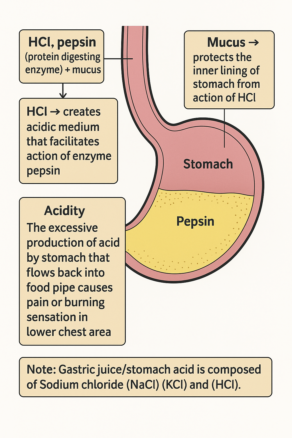
- Digestion in stomach taken care by gastric glands (present in walls of stomach)
Gastric glands release
- HCl, pepsin (protein digesting enzyme) + mucus.
- HCl → creates acidic medium that facilitates action of enzyme pepsin.
- Mucus → protects the inner lining of stomach from action of HCl.
- Acidity: The excessive production of acid by stomach that flows back into food pipe causes pain or burning sensation in lower chest area.
- Note: Gastric juice/ stomach acid is composed of Sodium chloride (NaCl) (KCl) and (HCl).
- From the stomach, food is slowly released into small intestine by sphincter muscle.
Small intestine:
- Small intestine is the longest part of alimentary canal fitted into compact space by extensive coiling (length ~7m).
- (Note: length of small intestine depends on food habits of animals.)
- Herbivores = long small intestine, since cellulose need to be digested.
- Carnivores = comparatively shorter small intenstine
- Small intestine is site of complete digestion of carbohydrates, proteins and fats.
- For digestion, it receives secretions from pancreas, liver.
- Food from stomach is acidic, this needs to make alkaline for the digestion.
- Bile juice from liver help to convert acidic → alkaline so that pancreatic enzymes can act and aid in digestion.
- Fats in small intestine are in the form of fat globules.
- It is difficult for enzymes to act.
- Bile salts break the fat globules into smaller globules and ↑ the efficiency of enzymes.
- Pancreas secrete pancreatic juice.
- Pancreatic juice contain enzyme trypsin for digesting proteins,
- lipase for breaking down emulsified fats.
- Walls of small intestine contain glands that secrete intestinal juice.
- The enzymes present in it convert
- proteins → aminoacids
- complex carbohydrates → glucose
- fats → fatty acids and glycerol
- Digested food is taken up by walls of intestine
- Inner lining of small intestine has numerous finger like projections called villi, that increase the surface area of absorption
- villi is supplied with blood vessels that takes the absorbed food into each and every cell of the body.
- This is used for 1) energy 2) Building new tissue 3) Repairing old tissues
Excretion:
- unabsorbed food is sent to large intestine, its wall absorb more water.
- Rest is removed from body via anus.
- Exit of waste material is by anal sphincter.
Dental caries
- It is also called as tooth decay.
- It is caused by softening of enamel and dentine.
- It starts when sugars bacteria act on sugars produce acid and soften/demineralise the enamel.
- Masses of bacteria with food particles stick to tooth to form dental plaque.
- Saliva cannot reach tooth surfaces to neutralise the acid, since plaque covers the tooth.
- Brushing teeth after eating removes plaque before bacteria produces acid.
- If untreated, microorganism invade pulp causing inflammation and infection.
RESPIRATION
- The food material taken in during the process of nutrition is used in cells to provide energy for various life process.
- Some use O₂ to breakdown into CO₂ and H₂O and other organism do not use O₂.
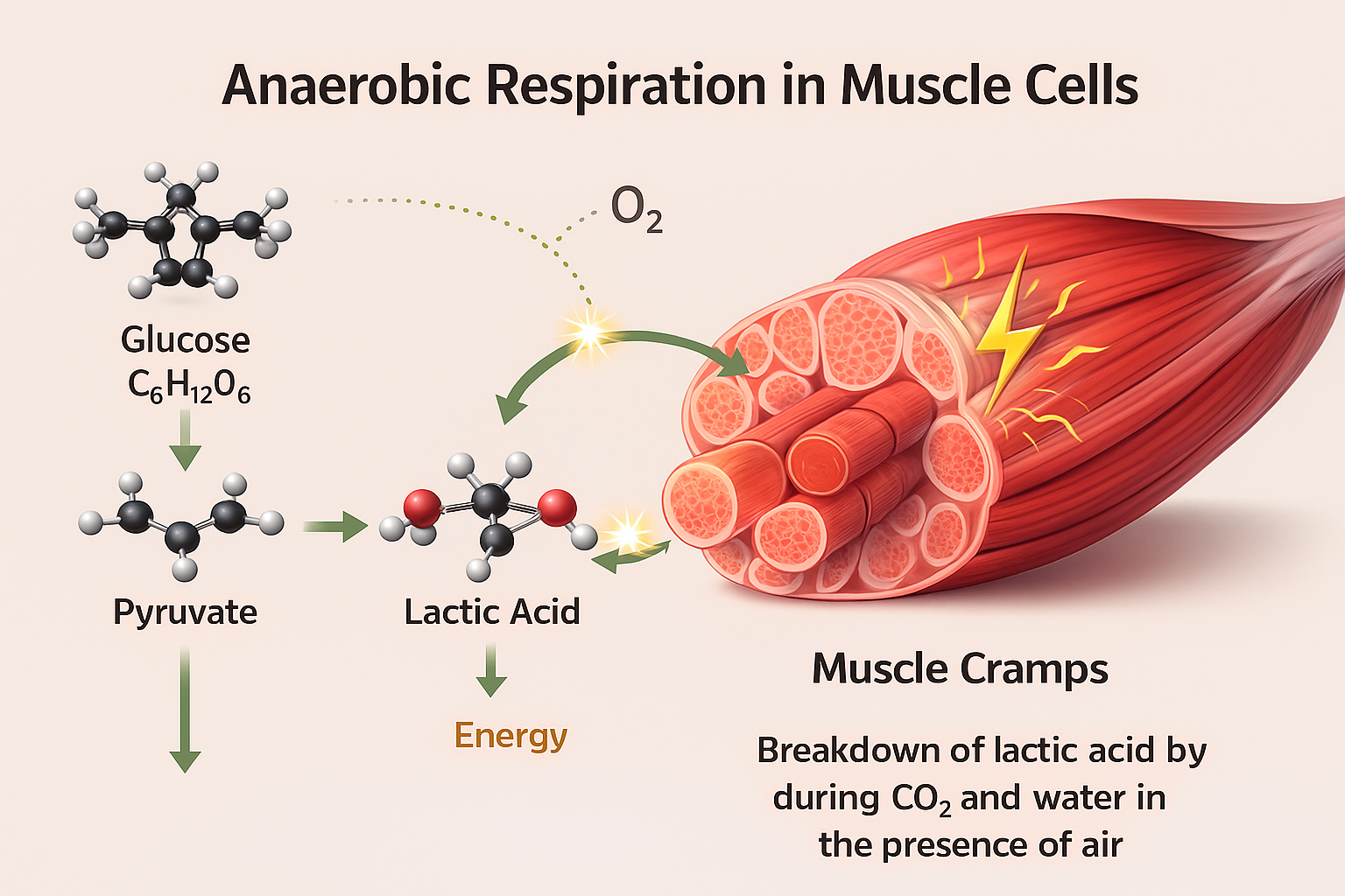
- In both cases, 1st step is breakdown of
- 6 carbon molecule of Glucose (C₆H₁₂O₆) into 3 carbon molecule called pyruvate.
- This process takes place in cytoplasm.
- In yeast during fermentation pyruvate is converted to ethanol (C₂H₅OH) and CO₂ in the absence of O₂. This is called anaerobic respiration.
- Breakdown of pyruvate using O₂ takes place in mitochondria.
- Here 3 carbon pyruvate breaks into CO₂ and water in the presence of air (O₂) and it is called aerobic respiration.
- Release of energy during aerobic respiration is more when compared to anaerobic respiration.
- Sometimes in our muscle cells, during lack of O₂, 3 carbon pyruvate is converted to lactic acid and energy.
- Buildup of lactic acid in muscle leads to cramps.
ATP
- Adenosine Tri phosphate.
- a) Energy released during cellular respiration is used to synthesise ATP.
- b) It is fuel to all the other activities of cell.
- c) ATP is energy currency for most cellular processes.
- d) The ATP is formed from ADP and inorganic phosphate.
- ADP + P + Energy → ADP–P = ATP
- The endothermic process that takes place in cell use this ATP.
- When the terminal phosphate link in ATP is broken using water, energy ≈ to 30.5 kJ/mol is released.
- ATP is used in cells for contraction of muscles, protein synthesis, conduction of nerve impulses and other activities.
Aerobic Respiration:
conditions required:
- Aerobic = O₂ dependent
- Sufficient intake of O₂
- plants → Gaseous exchange takes place through stomata
- Larger intercellular spaces are present for the proper diffusion of O₂ and CO₂
How intercellular spaces helps in plant respiration?
- Cells have large intercellular space for the gaseous exchange.
- The air need to go in and come out of the cell.
- Direction of diffusion depends on
- i) Environmental conditions
- ii) Plant requirements.
- At night, there is no photosynthesis so CO₂ elimination takes place.
- During day, since photosynthesis takes place O₂ release is major event.
O₂ absorption in terrestrial organism.
Terrestrial organism breathe the O₂ from the atmosphere.
Since air contain more O₂, terrestrial organism do not have to breathe faster.
Terrestrial organism breathe through
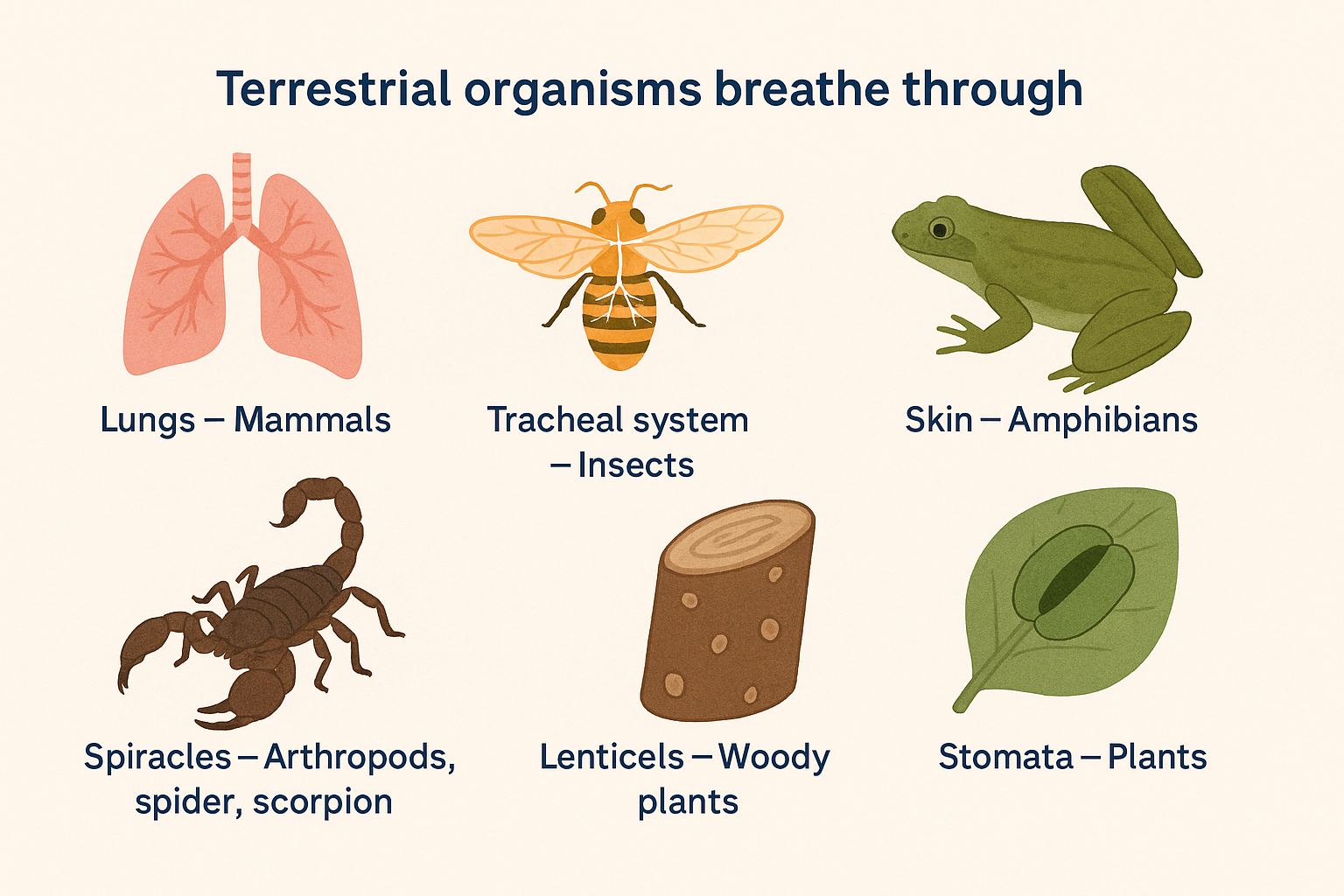
- Lungs – Mammals
- Tracheal system – Insects
- Skin – amphibians
- Spiracles – Arthropods, spider, scorpion
- Lenticels – woody plants
- Stomata – plants
- Gaseous exchange takes place through the above organs in various organism. Since all of them are delicate structures, they are placed inside the body.
- Also, there is various mechanism for air moving in and out of the area and O₂ is absorbed.
Aquatic animals
- They breathe through gills.
- The amount of dissolved O₂ is comparatively low than atmospheric O₂, so the rate of respiration/ breathing in aquatic organism is much faster.
Human Respiration:
- The air is taken into the body through Nostrils.
- Nostrils have fine hair, it filters the air passing through it.
- Nostril passage is lined with Mucus (viscous substance) that filters the air from dust & dirt.
- (Mucus – slippery viscous substance produced by mucus membrane. It has water, glycoprotein, salts, cells etc. Its function is protective barrier lining the surface of organs.)
- Air passes into throat and then into the lungs.
- Trachea – wind pipe extends from larynx (voice box to bronchi)
- Rings of cartilage = Ring of cartilagenous tissues found in Trachea (windpipe)
- Cartilage = tough flexible connective tissue found in joint, ears, nose.
- Rings of cartilage are present in throat. It ensures the unimpeded (not obstructed)/ uninterrupted flow of air into and out of lungs during breathing.
- Within the lungs, passage divides into smaller tubes and finally terminate into balloon like structure called alveoli.
- Alveoli provide surface for gaseous exchange.
- Walls of alveoli have extensive blood vessels.
- The diaphragm moves down and ribcage expands as we breathe in. This creates more space in chest and so air rushes into lungs.
- 10. Blood brings CO₂ from rest of body & releases into alveoli.
- O₂ in the alveolar air is taken up by blood in alveolar blood vessels and is transported to all cells of the body.
- During breathing cycle there is always a residual volume of air, so that there is enough time for O₂ to be absorbed and CO₂ to be released.
Role of Respiratory Pigments
- When the size of animal is large, diffusion pressure is not enough for carrying O₂ to all the cells of the body.
- Here comes the respiratory pigment. They take up O₂ from air in lungs and carry it to tissues that are O₂ deficient.
- In Humans, respiratory pigment is Hb (Haemoglobin). It is present in RBC and has high affinity for O₂.
- CO₂ is more soluble in water, hence transported in dissolved form in our blood.
TRANSPORTATION
- Blood is a fluid connective tissue.
- It consists of fluid medium called plasma, in this cells are suspended.
- Plasma transports food, CO₂, nitrogenous waste in dissolved form.
- Apart from this, salts are also transported by the blood.
- We need pumping organ to push the blood to various organs of our body and network of tubes to reach all the tissues.
Pump – HEART
- Heart is a muscular organ. It is size of our fist.
- The heart has different chambers to carry the O₂ rich blood and CO₂ containing blood (deoxygenated blood).
- CO₂ rich blood has to reach the lungs to be removed.
- O₂ rich blood from lungs is brought back to heart so that it can be pumped to the rest of our body.
Step by step process
- Oxygenated blood from lungs comes to left atrium (upper chamber – left side)
- It is relaxed when blood is collected
- Left atrium contracts and blood is flowed to left ventricle (It is now expanded)
- When left ventricle contracts, blood is pumped out to the body.
- Deoxygenated blood comes from body to right atrium (upper chamber – right) (expands)
- Right atrium contracts, blood is flowed/ transferred to right ventricle
- Right ventricle contracts and this blood is pumped into lungs for oxygenation.
- Ventricles have to pump the blood into various organs, they have thick muscular valves.
- Valves are present to ensure that blood do not backflow when atria/ventricles contract.
Difference between Auricles and Ventricles
| S.No | Auricles (Atria) | Ventricles |
|---|---|---|
| 1 | Two upper chambers of the heart | Two lower chambers of the heart |
| 2 | Types: Right atrium & Left atrium | Types: Right ventricle & Left ventricle |
| 3 | Thin-walled chambers | Thick and muscular walls |
| 4 | Receive blood: from body (right atrium), from lungs (left atrium) | Pump blood: to lungs (right ventricle), to the body (left ventricle) |
| 5 | Primary function: collect blood and pump it into ventricles | Primary function: pump blood to lungs/body |
| 6 | Contain atrioventricular valves: Tricuspid (right), Mitral (left) | Contain atrioventricular + semilunar valves: Pulmonary (right), Aortic (left) |
Types Of Circulation:
| Feature | Double Circulation | Pulmonary Circulation | Systemic Circulation |
|---|---|---|---|
| Definition | Circulation of blood through two separate pathways in the body. | Circulation of blood between the heart and lungs. | Circulation of blood between the heart and all body parts (except lungs). |
| Pathway Count | Has two circuits: pulmonary + systemic. | One circuit: heart → lungs → heart. | One circuit: heart → body → heart. |
| Purpose | Ensures complete separation of oxygenated and deoxygenated blood. | Oxygenates blood and removes CO₂. | Supplies oxygenated blood to the entire body and collects CO₂ & wastes. |
| Blood Type Carried | Involves both oxygenated and deoxygenated blood. | Carries deoxygenated blood to lungs and returns oxygenated blood to heart. | Carries oxygenated blood from heart to body and returns deoxygenated blood to heart. |
| Main Function | Maintains efficient oxygen supply for high metabolism. | Gas exchange: oxygen uptake & CO₂ removal. | Nutrient delivery, waste removal, and maintaining body functions. |
| Involving Heart Chambers | Uses all four chambers of the heart. | Right ventricle → Lungs → Left atrium. | Left ventricle → Body → Right atrium. |
Blood pressure
- The force that blood exerts on the walls of blood vessel is blood pressure.
- Blood Pressure is measured using Sphygmomanometer. Blood Pressure → (Phase of heartbeat)
- Heart beat = one complete cycle of systole and diastole.
- Systole = Heart muscle contract, pumps blood from chambers into arteries.
- Diastole = Heart muscle relax, allow chamber to fill with blood.
- Systolic pressure = pressure of blood inside arteries during ventricular systole (contraction). Normal – 120 mm of Hg.
- Diastole pressure = pressure in artery during ventricular diastole (relaxation) is diastolic pressure. Normal – 80 mm of Hg.
| Feature | Arteries | Veins | Capillaries |
|---|---|---|---|
| 1. Function | Carry blood away from the heart to body organs | Collect blood from organs and bring it back to the heart | Connect arteries and veins; allow exchange of materials |
| 2. Wall Thickness | Thick and elastic walls | Thin walls | Walls are one-cell thick |
| 3. Type of Blood Carried | Usually carry oxygenated blood (except pulmonary artery) | Usually carry deoxygenated blood (except pulmonary vein) | Carry mixed blood for exchange of gases and nutrients |
| 4. Location | Located deep inside the body | Often close to skin surface | Present within organs and tissues |
| 5. Valves | No valves | Have valves to prevent backflow | No valves |
| 6. Lumen (Opening) | Narrow opening | Wider opening | Very narrow, just enough for single blood cells |
| 7. Speed of Blood Flow | Blood flows quickly | Blood flows slowly | Blood flow is very slow to allow exchange |
| 8. Pressure | High pressure (blood pumped directly by heart) | Low pressure | Lowest pressure |
| 9. Additional Notes | — | — | Arteries branch → arterioles → capillaries → venules → veins |
Function of platelets:
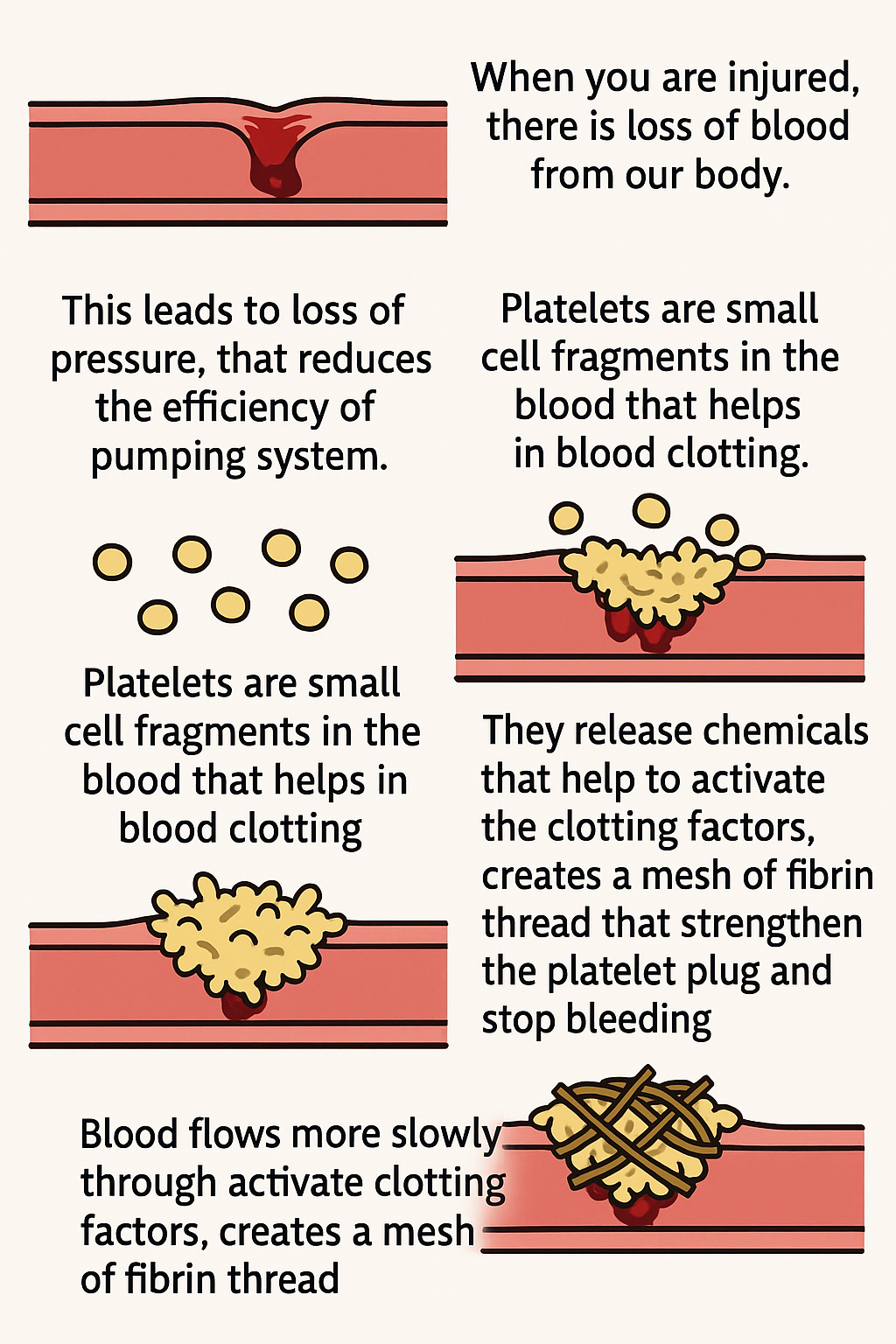
- When you are injured, there is loss of blood from our body.
- This leads to loss of pressure, that reduces the efficiency of pumping system.
- Platelets are small cell fragments in the blood that helps in blood clotting.
- After injury, platelets gather at site of wound. and stick together to form plug.
- They release chemicals that help to activate the clotting factors, creates a mesh of fibrin thread that strengthen the platelet plug and stop bleeding.
Lymph
- It is a colourless fluid containing less proteins and involved in transportation.
- It is also called as tissue fluid.
- Through the pores in the capillaries, some amount of plasma, proteins, blood cells escape into intercellular space in tissues to form tissue fluid/lymph.
- Lymph drains into lymphatic capillaries, they join to form lymph vessels and finally open into larger veins.
- Function:Lymph carries digested and absorbed fat from intestine and drains excess fluid from extra cellular space back into blood.
Transportation in plants
- Plants make their own food through leaves by photosynthesis process.
- Soil is the nearest and richest source of raw materials like N₂, phosphorus and other nutrients.
- Roots help in the absorption of these nutrients to plants.
- When the distance between the soil contacting organ (root) and chlorophyll containing organ (leaves) is small, energy and raw materials can easily diffuse to all parts of body.
- But when the distance is large, a proper transportation is required for the transport of all nutrients.
- Plant transport system will move
- Energy – stored in leaves
- Raw materials – from roots
- Xylem = It moves water and minerals obtained from soil.
- Phloem = Transports product of photosynthesis from leaves to other parts of plant.
TRANSPORT OF WATER
- In xylem tissues vessels and tracheids of the root, stem, leaves are interconnected to form continuous system of water conducting channels to reach all plant parts.
- In root, cell in contact with soil take up the ions.
- This creates difference in concentration of ions between root and soil.
- Water moves from soil into root to eliminate this difference.
- There is steady movement of water into root xylem, creating a column of water that is steadily pushed upwards.
Another method:
Evaporation of water molecules from cells of leaf creates a suction, that pulls water from xylem cells of root.
TRANSPIRATION
- Loss of water in the form of vapour from aerial parts of plants is known as transpiration.
- Transpiration helps in absorption and upward movement of water and minerals dissolved in it from root to leaves.
- ↑ the osmotic pressure of tissue causing H₂O to move into it.
- This pressure moves the material in phloem to tissues that has less pressure.
- This happens according to plant needs. Ex: During spring, sugar stored in root/stem transported to buds that needs energy to grow.
| S.No | Xylem | Phloem |
|---|---|---|
| 1. Function | Transports water and minerals from roots to all parts of the plant. | Transports food (glucose/sucrose) from leaves to all parts of the plant. |
| 2. Direction of Flow | Movement is unidirectional (only upward). | Movement is bidirectional (upward & downward). |
| 3. Role | Helps in water conduction and provides mechanical support. | Helps in translocation of food prepared during photosynthesis. |
| 4. Composition | Made up of tracheids, vessels, xylem fibres, xylem parenchyma. | Made up of sieve tubes, companion cells, phloem fibres, phloem parenchyma. |
| 5. Cell Type | Mainly contains dead cells (tracheids & vessels). | Mainly contains living cells (sieve tubes & companion cells). |
| 6. Wall Thickness | Cells have thick, lignified walls. | Cell walls are thin and not lignified. |
| 7. Energy Requirement | No energy required, passive transport. | Energy required (ATP) for food transport. |
| 8. Location | Located in the inner portion of vascular bundles. | Located in the outer portion of vascular bundles. |
EXCRETION
- The biological process involved in the removal of those harmful metabolic wastes from the body is called excretion.
- Unicellular organism – Remove waste by simple diffusion.
- Multicellular organism – Use specialised organs to remove waste.
Human beings
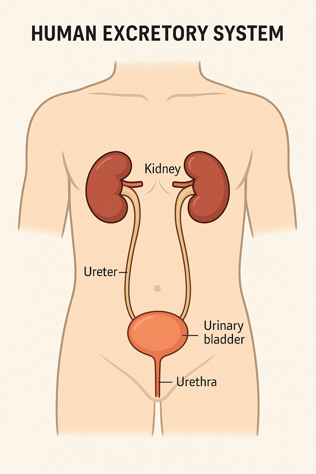
- Excretory system of humans has:
- a) pair of kidney
- b) pair of ureters
- c) urinary bladder
- d) urethra
Location: Kidneys located in the abdomen, one on either side of the backbone.
- Urine produced in kidneys
- It passes through ureters
- It is finally stored in urinary bladder
- Through urethra, urine is expelled
Urine:
- Composition: Urine consist of nitrogenous waste such as urea / uric acid.
- These are removed from blood in the kidney.
Excreatory system working principle:
- Like lungs, kidneys also have cluster of thin walled blood capillaries.
- Each capillary cluster in the kidney is with cup shape end of tube (Bowman’s capsule). It collects the filtered urine.
- Each kidney has large number of filtration unit called nephrons. These are packed close together.
- Glucose, Aminoacids ,Salts, Major amount of water → All these are selectively reabsorbed as urine flows along the tube.
- Amount of water reabsorbed depends on
- a) How much excess water in the body
- b) How much of dissolved waste is to be excreted.
- Urine forming in the kidney enters the ureter.
- Ureter connects the kidney with the urinary bladder.
- Urine is stored in the urinary bladder.
- Bladder is muscular and it is under nervous control.
- The pressure of the expanded urinary bladder leads to urge and then urine is passed through urethra.
- Since it is under nervous control, we usually control the urge to urinate.
ARTIFICIAL KIDNEY
(HEMODIALYSIS)
- The function of kidneys is affected by infections, injury, restricted blood supply.
- This can lead to accumulation of poisonous waste in the body.
- Artificial kidney is used to remove nitrogenous waste products from blood through dialysis.
Steps:
- Artificial kidneys contains number of tubes with semipermeable lining.
- It is suspended in tank filled with dialysing fluid.
- This fluid has same osmotic pressure as blood.
- It is devoid of nitrogenous waste.
- Blood is passed through these tubes.
- During this process, waste products from blood pass into dialysing fluid by diffusion.
- Purified blood is pumped back to kidney.
- In normal human kidneys, initial filtrate is 180 L daily but we excrete only about 1 or 2 litre a day.
- The rest is reabsorbed in kidney tubules.
- But in dialysis, there is no reabsorption of the filtrate takes place.
Excretion in plants
- Plants get rid of excess water by transpiration.
- Leaves that fall off from plants contain dead cells and plant waste products are stored in cellular vacuoles.
- Waste products stored in leaves fall off.
- Resin, gums are waste products stored in old xylem. Plants excrete some waste substances into soil around them.